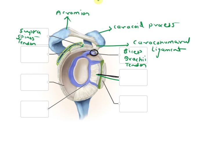
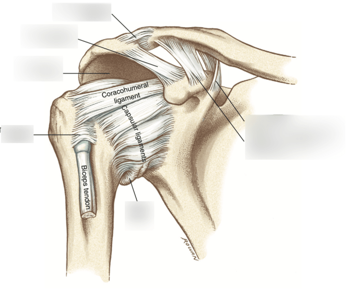
Correctly identify the following anatomical parts of the glenohumeral joint – Correctly identifying the anatomical parts of the glenohumeral joint is essential for understanding its structure and function. This guide will provide a comprehensive overview of the glenoid cavity, humeral head, ligaments, muscles, and nerves that make up this complex joint, offering valuable insights into its mechanics and clinical implications.
Glenohumeral Joint Anatomy

The glenohumeral joint, also known as the shoulder joint, is a ball-and-socket joint that connects the humerus (upper arm bone) to the scapula (shoulder blade). It is a complex joint that allows for a wide range of motion, including flexion, extension, abduction, adduction, and rotation.
Glenoid Cavity

The glenoid cavity is a shallow, concave socket located on the lateral aspect of the scapula. It is lined with a layer of articular cartilage that provides a smooth surface for the humeral head to glide against. The glenoid labrum is a fibrocartilaginous ring that surrounds the glenoid cavity and helps to deepen it.
The glenoid labrum also helps to stabilize the shoulder joint by preventing the humeral head from dislocating.
Humeral Head: Correctly Identify The Following Anatomical Parts Of The Glenohumeral Joint

The humeral head is the rounded end of the humerus that fits into the glenoid cavity. It is covered with a layer of articular cartilage that reduces friction and wear. The shape of the humeral head allows for a wide range of motion, including flexion, extension, abduction, adduction, and rotation.
Ligaments
The glenohumeral joint is stabilized by a number of ligaments, including the anterior glenohumeral ligament, the posterior glenohumeral ligament, the inferior glenohumeral ligament, and the superior glenohumeral ligament. These ligaments work together to prevent excessive movement and dislocation of the shoulder joint.
Muscles
The glenohumeral joint is surrounded by a number of muscles that allow for movement of the shoulder. These muscles include the deltoid muscle, the supraspinatus muscle, the infraspinatus muscle, the teres minor muscle, and the subscapularis muscle. These muscles work together to produce coordinated movement of the shoulder joint.
Nerves

The glenohumeral joint is innervated by a number of nerves, including the axillary nerve, the suprascapular nerve, and the subscapular nerve. These nerves transmit sensory and motor signals to and from the shoulder joint. Damage to these nerves can result in loss of sensation or function in the shoulder joint.
Popular Questions
What is the glenoid cavity?
The glenoid cavity is a shallow, concave depression on the lateral surface of the scapula that forms the socket of the shoulder joint.
What is the role of the glenoid labrum?
The glenoid labrum is a fibrocartilaginous rim that surrounds the glenoid cavity, deepening it and providing stability to the shoulder joint.
What are the major ligaments of the glenohumeral joint?
The major ligaments of the glenohumeral joint include the superior glenohumeral ligament, middle glenohumeral ligament, and inferior glenohumeral ligament, which work together to stabilize the joint and prevent excessive movement.